Automate your analysis
A Central Resource
Analysis and Storage
- Secure Storage
Images and results securely stored in the cloud on AWS. Collaborate with other institutions. Images are private by default. No PHI. Data encrypted at rest and in motion.
- Central Analysis
A centralized image analysis core that can be accessed from anywhere. Full automation minimizes human errors. Built on the robust ANTsX ecosystem. Finally, a way to compare across studies.
- Save Money and Time
Dramatically reduce time spent performing manual segmentations and analysis. No need to worry about fixing broken code after a software update.
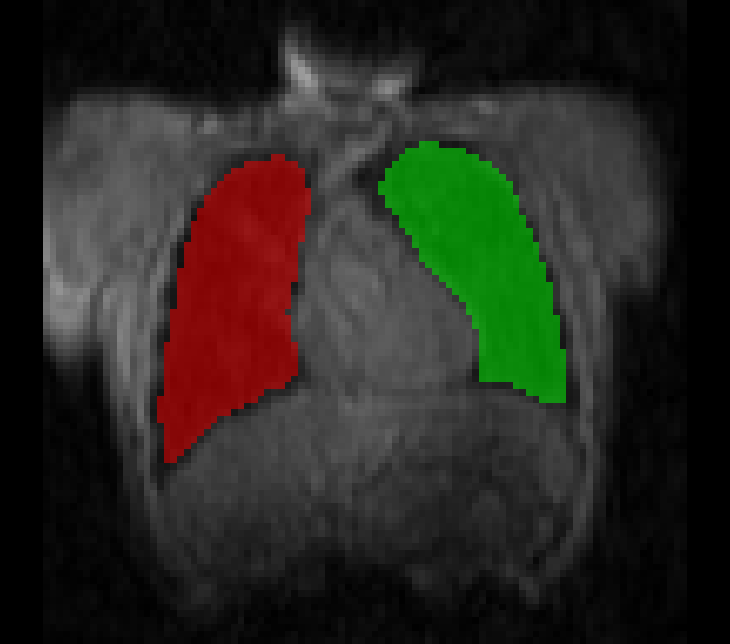
Thoracic Segmentation
Access to automation
- Save time
Because you have better things to do than perform manual segmentations. Learn more about the technique.
- Anatomic MRI
Automated thoracic segmentation of MR images
- Customize
Submit training data to have the model custom trained to work with your images
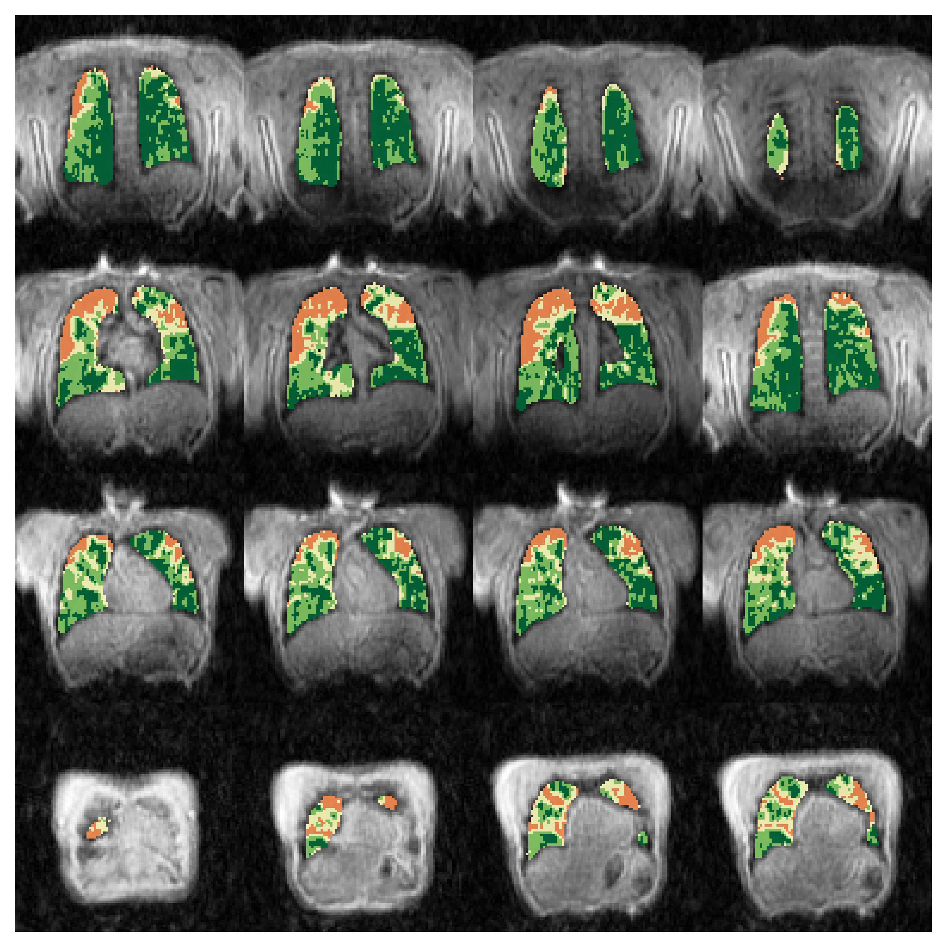
VDP Analysis
A unified analysis
- Deep learning
Ventilation analysis performed using ANTsPyNet's El Bicho. Learn more about image vs histogram based semantic segmentation.
- Our mask or yours
Provide an anatomic image and we can perform the ventilation analysis automagically within the area defined by the thoracic cavity or submit a manual mask to constrain the analysis.
- Quantification
Export ventilation quantification reports to csv.
ADC
Diffusion weighted MRI
- Microstructure
Measure lung microstructure with 3D diffusion weighted MRI.
- Plots
Visual plots as well as quantitative measurements.
- The Equation
You can provide a mask or we can create one. ADC calculated using S_b = S_0*exp(-ADC*bvalue)
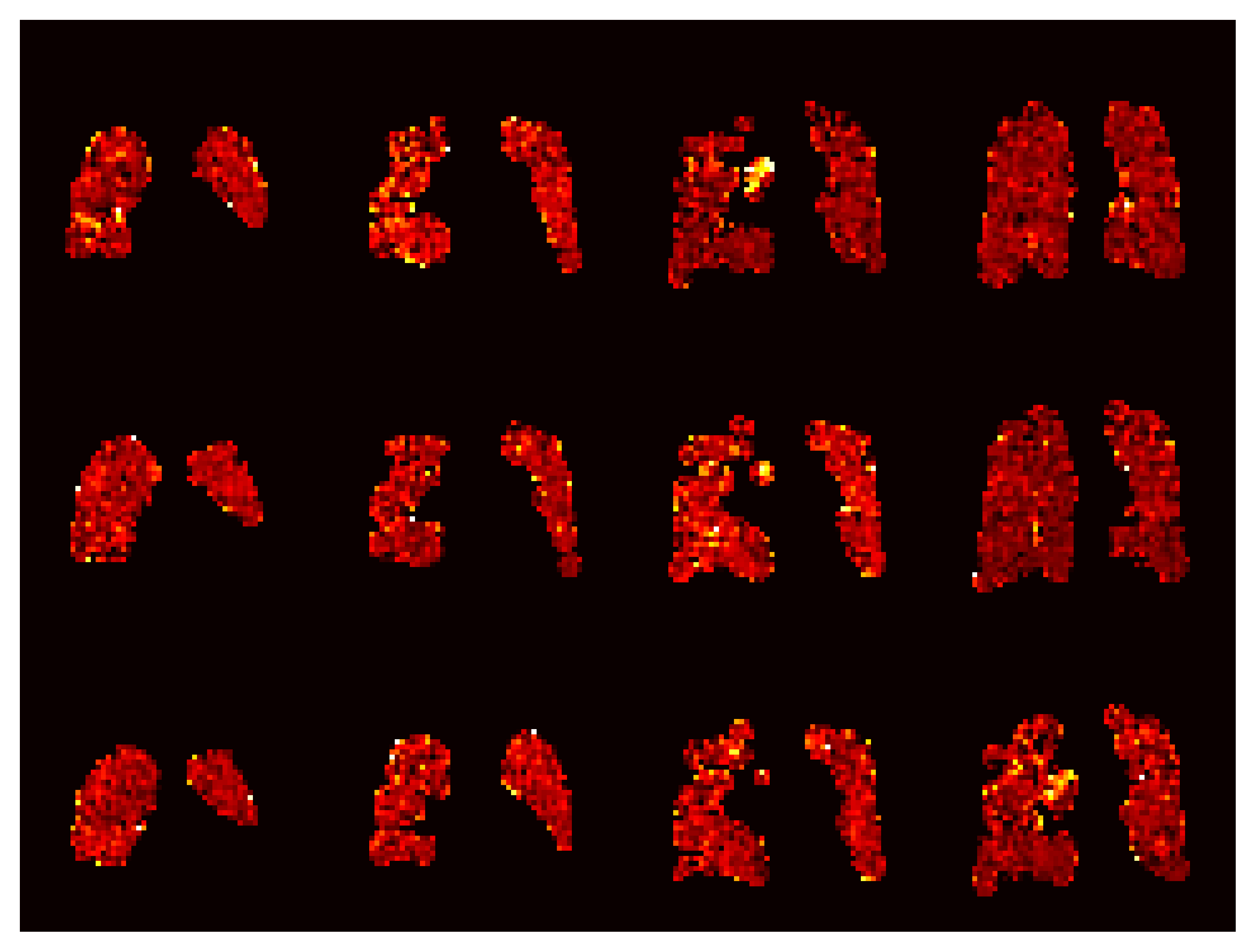
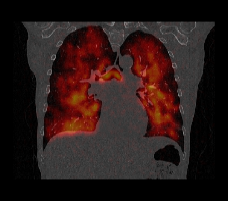
Image Registration
Made Simple
- 3D registration of MR to CT
Map ventilation and segmentation images to CT space.
- No prior experience needed
We handle the details.
- Expand your analysis
Perform regional comparisons to CT.